Misinterpretastasis.

Understanding the ‘theory and interpretation’ of metastasis.
The standard medicine tells us that a primary cancer has spread (‘mets’) to a secondary and tertiary organ. This is commonly known as the theory of metastasis. Yet, even the prevalent mechanisms are mere hypotheses.
For those that have been following these blog posts | case studies – you’re already quite familiar with the notion that a biological conflict will impact us at the level of the consciousness, brain and organ.
The organ manifestation has been discussed many a time, in various posts.
What we haven’t spent much time with is the impact the conflict has on the cerebral or brain level.
The brain mediates the DHS (biological conflict) between the psyche and the target organ.
The biological conflict initially impacts the awareness on the (sub)conscious level, then alights a predetermined relay in the brain, as a short-circuit or bio-electrical imprint (visible on a brain scan) – the location of which is exclusively based upon the conflict’s unique nature or theme.
A natural emergency adaptation or survival program, with the accompanying organic and functional tissue change is set into motion in the corresponding organ system.
This bio-electrical imprint, officially known as a hamerscher herde, appears on a brain CT scan (computerized tomography) as a target configuration or series of concentric rings – like the ripple effect we observe when a stone hits the water on a lake. This bio-electrical imprint is actually a three dimensional sphere, much like a fireworks display – yet since a brain CT scan is sectioned in layers or slices the imprint appears two dimensional.
A highly trained eye can ascertain one’s entire medical story from a brain scan. When the bio-electrical imprint or target ring is fresh with very clear and sharp lines – the DHS or biological conflict is new and active. Upon resolution of the conflict, the target ring configuration begins to break up. The once clearly defined rings will now appear to be intermeshed with what resembles the spokes on a bicycle wheel in appearance. As the healing phase unfolds on the organ level it also unfolds on the cerebral or brain level. The ring formation becomes less and less defined.
What relevance does this have to ‘metastasis’?
When we look at a client’s symptomatic presentation or diagnosis we observe a hamerscher herde (in the exact stage of radiological progression) for each and every symptom or diagnosis (in the exact stage of symptomatic progression.) There is no way around this fact.
Let me serve an example from personal experience. In 2009, my mother was diagnosed with stage 3b ‘metastatic’ adenocarcinoma.
A single life event can impact us in symphony. One traumatic event may have multiple facets.
In my mom’s scenario a single life event was perceived by her psyche as a territorial aggression, translated into a bronchial program (*bronchial carcinoma), while heading into a dangerous situation translated into a branchial arch program (mistakenly identified as metastatic lymph nodes.)
In this scenario there was also a territorial anger over property (mistakenly identified as bile duct involvement.)
Above, I said my mom was diagnosed with stage 3b ‘metastatic’ adenocarcinoma. That diagnosis was in fact wrong. Rather, she was expressing a bronchial carcinoma – different part of the brain, different brain relay) … different conflict. Fine needle biopsies are not 100% accurate as the needle often goes through multiple tissue types and therefore multiple germ layers.
In 2004, my parents were going through a less than amicable divorce. It had gotten to the point where one of them was going to have to move out of the house – yet neither of them would budge. This territorial stalemate went on for number of months.
My mothers attorney suggested that, through a court order, my father be physically removed from the house via the sheriff. The day the sheriff was to come to come, unannounced, to remove my father from his home – my mother called me in the morning telling me she could not sleep the night before and that she could not go through with it. She could not have my father forcibly removed from the house.
This territorial aggression was her dhs (biological conflict) – feeling aggressed towards or unsafe within her home, within her territory.
Ironically, it would have been impossible for my mom to have experienced a masculine territorial conflict even a few years earlier. One way a woman can experience a masculine conflict is if they are biologically no longer female, meaning postmenopausal, which my mom was.
That territorial stand-off was responsible for the bronchial program.
At the same, time the thought for my mother of having to leave the house, where would she go, how would she survive – was a concurrent biological conflict shock of heading into a dangerous situation. This was responsible for the branchial arch program (mistaken as ‘metastatic’ spread to the mediastinal lymph nodes.)
Finally, another concurrent biological conflict involved a territorial anger over who will remain in the house which initiated the bile duct program which was misinterpreted as bile duct involvement.
In December 2008, my mother came to terms with all of this and all three biological programs, active for five years went into resolution in “symphony” and she was diagnosed with lung cancer that had ‘metastasized’ to the lymph nodes and bile ducts.
On her CT, I observed a healing phase hamerscher herde evident in the cerebral cortex, in the exact relay for the bronchial mucosa and bronchial musculature.
On her CT, I observed a healing phase hamerscher herde evident in the cerebral cortex, in the exact relay for the “lymph nodes.”
On her CT, I observed a healing phase hamerscher herde evident in the cortex once again, in the exact relay for the bile ducts.
There was no stage ‘3b’ metastatic adenocarcinoma, rather the ‘metastasis’ was explained by concurrent biological conflicts resolving in symphony.
If you take the time to understand The Five Biological Laws of GNM – you’ll understand that ‘metastasis’ as it is presently explained by conventional thought is a misinterpretation.
Dr. Hamer does not dispute the existence of secondary and tertiary cancers – but the notion of metastasis from a primary site becomes a questionable fairytale in light of the above (and other) observations. More accurate is that metastases are the result of concurrent or sequential biological conflicts.
I have observed the above over and over and over with not only metastases but with syndromes that have multiple symptoms, read that multiple conflicts/biological programs running … Lyme Disease, AIDS, Guillain Barre Syndrome et al. In each case, the clients history and brain scan demonstrated they experienced the exact biological conflict for each symptom or ‘metastatic’ location.
There is no way around this.
There are no exceptions.
There are numerable other factors that dispute the present concept of metastasis.
All organs in the body originate from three basic tissue types, called germ layers. Associated with each type of germ layer are microbes (fungus, bacteria etc.) that assist with healing. Primary germ layers cannot transform themselves after conception. In other words, apples cannot become oranges and oranges cannot become limes.
Endodermal tissue is controlled by the brainstem.
Mesodermal tissue is controlled by the cerebral medulla.
Ectodermal by the cerebral cortex.
What Dr. Hamer discovered is that each tissue type (as well as behavior) is controlled by a very specific part of the brain. It would be implausible for a brain stem controlled, endodermal cancer cell that demonstrates cell(+) augmentation behavior and mycobacteria expression in healing phases (glandular breast, prostate, e.g.) to magically spread and also transform to a medulla controlled, mesodermal cancer cell that now demonstrates cell (-) ulceration behavior and bacterial expression in healing phases (bone, lymph nodes, e.g.)
If a different brain relay, different embryonic germ layer and different microbial expression is responsible for and associated with the secondary cancer, how can this possibly be attributed to ‘metastasis’?
Metastasis in the conventional sense cannot exist in light of The Five Biological Laws. There is a fly in the metastatic ointment.
This and other factors will be discussed in a future blog, but before concluding this one I want to touch upon the present scientific method utilized today for observing metastasis.
When tumor cells metastasize, the new tumor is called a secondary or metastatic tumor, and its cells are (*said to be) similar to those in the original or primary tumor. This means, for example, that, if breast cancer metastasizes to the lungs, the secondary tumor is (*said to be) made up of abnormal breast cells, not of abnormal lung cells. The tumor in. the lung is then called metastatic breast cancer, not lung cancer. (Wikipedia, *with notation.)
Anne Mcilroy, in her 2006 expose entitled The End of chemo? One Magic Pill May Hold Answer references Dr. Vincent Giguère’s oncology research where his team observed that “Breast-cancer cells, for example, often move to the bones. This is quite a feat, since they first have to morph from breast cells into bone cells.” He and his colleagues are trying to figure out how they do it – what makes them different from the cells in the bulk of the tumour, which stay in the breast?”
Heidi Ledford, in her April 19, 2011 Cancer Theory Faces Doubts. “A leading explanation for how cancer migrates falls short on clinical evidence. It can’t be easy to stand in front of hundreds of colleagues and tell them that ten years of research has led them in the wrong direction. But at the annual meeting of the American Association for Cancer Research (AACR) earlier this month, pathologist David Tarin did just that, by challenging a leading hypothesis on how cancers metastasize. Tarin, from the University of California, San Diego, questioned the idea that cancer cells break free and migrate to new sites in the body. But Tarin, along with some other cancer biologists, argues that no one has seen the process in action in human cancers.”
John Pawelek, a researcher in the Department of Dermatology at Yale School of Medicine and Yale Cancer Center, speaking about his “Fusion Theory” of metastasis (May 2008 Nature Reviews Cancer) states …“Although we know a vast amount about cancer, how a cancer cell becomes metastatic still remains a mystery.”
So how do histopathologists determine the ‘metastatic’ origin of a secondary or tertiary cancer?
In a word … indirectly.
The laboratory protocols are so complex, inverted and circular, that the old adage of not being able to see the forest for the trees applies here in spades. The testing is always by indirect means.
In the most simple explanation, what oncology labs (immunohistochemistry) are looking for, through a series of complex and indirect biochemical staining procedures – is the presence of markers or antigens found in or on the cancer cells that are associated with a particular tumor type. This method is not designed to identify the presence of metastasizing cancer cells, rather surface proteins that are associated with a primary tumor.
Why is this significant?
Metastasizing cancer cells are not being observed – only harmless surface proteins associated with a primary tumor are. Even the standard medicine admits they are in the dark when it comes to an understanding of all these proteins, why some react and others do not.
One hypothesis is that these proteins are a natural part of the cells biochemical expression during certain biological conflict active and healing phases.
An example of this is seen in prostatic adenocarcinoma. As the patient enters the healing phase, mycobacteria will degrade the cell growth that built up during conflict activity. The breakdown releases proteins (NOT metastatic cancer cells) from the original tumor which may end up “in-the-mix” at the biopsied secondary site.
It is not unheard of for a needle biopsy diagnosis to be wrong as multiple embryologic germ layers (and therefore surface proteins) are accessed during the procedure.
Today’s methodology makes the indirect conclusion that the presence of these surface proteins, typical of the primary tumor are evidence of ‘metastasis.’ That’s one big leap!
The relevance of cancer biomarkers (tumor markers) comes under scrutiny for reasons of likewise application and interpretation (that’s another blog, for another day.)

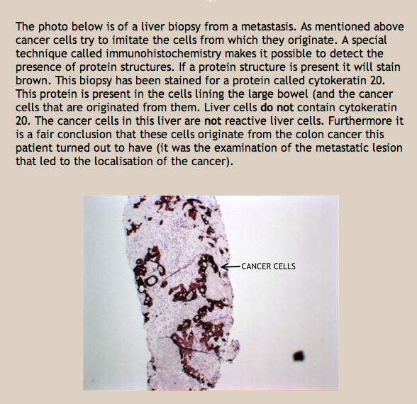
What the above photo taken off the internet is showing is NOT colon cancer cells that have metastasized to the liver but rather surface proteins from the colon cancer cells appearing in the liver.
A final observation. In order to understand why metastases may appear we first need to appreciate the Second Biological Law of German New Medicine (and it’s interplay with agents such as chemotherapy) – simply stated, the law of two phases. Every disease unfolds with a conflict active and healing phase (assuming a conflict resolution has been reached.)
The conflict active phase is dominated by what is termed sympathicotonia – the cold phase of the sympathetic nervous system, while the healing phase is dominated by what is termed vagotonia – the warm phase of the parasympathetic nervous system.
As we have discussed, some biological programs (cancers) show tissue augmentation during the conflict active phase, while others only during the resolution or healing phase.
Let’s look at an individual with a sarcoma of the humerus (bone cancer of the arm) as background. The individual had been through chemotherapy and everything was looking good. A couple of months later the sarcoma returned and was now, according to the oncologist, growing rapidly and with a great aggressiveness. A sarcoma program (bone cancer) will demonstrate cell loss during the conflict active or sympathicotonic phase of the biological program.
If the conflict was still active when the chemotherapy was administered – the cancer will “return” six to eight weeks after the chemo has left the body. In actuality, nothing is “returning”, rather the expression of the natural biological program was affected by the treatment, but with an adverse complicating twist … chemotherapy is sympathicotonic, so the treatment will interrupt or suspend the healing phase by inducing a deeper state of sympathicotonia. If the sarcoma conflict was still active (sympathicotonia) when the chemotherapy was initiated, the chemotherapy will deepen the conflict active phase.
When the chemotherapeutic agents leave the body, the healing phase recommences but with a rebound effect due to this amplified sympathicotonia. As the biological program starts up again with an intensified expression – this is interpreted by traditional oncology as an “aggressive” return of the cancer. Sadly, we see this over and over again.
This observation applies to radiation therapy as well. It should be noted that steroids are often administered during chemotherapy – this is a double whammy (I know, I know – not very technical) as the steroid has the same effect as described above.
So the correlative observation here relative to metastasis is if there are very minor (subclinical), let’s call them trace biological programs running (undetectable via todays diagnostic
technology) at the time chemotherapy is administered for a primary (and the yet only identifiable cancer) – once the chemotherapeutic agent has left the body the same rebound effect (amplified sympathicotonia) will occur … making these very minor, trace biological programs – figuratively explode into expression and now become detectable via todays diagnostic technology.
Cornell Chronicle. By Renee Twombly | November 16, 2015. For more than a decade, many researchers have believed that a biological process that transforms the shape of cells that line cavities, organs and blood vessels in the body was necessary for metastasis. Epithelial to mesenchymal transition, or EMT, strips away the cells’ ability to hold on tightly to their neighbors, allowing them to migrate throughout the body. In their study published Nov. 11 in Nature, Weill Cornell Medicine investigators discovered that while EMT occurred in a small number of primary breast tumor cells, they were not involved in cancer metastasis. What’s more, metastasis was derived from non-EMT cancer cells, contradicting the common theory about how cancers spread. Strikingly, the EMT status changed with the addition of chemotherapy.
This is just a personal observation on my behalf. But when we look at the prevalence of chemotherapy in treatment today (nearly 100%) it becomes a working hypothesis that needs to be deeply studied.
This blog is an educational only blog. The information and services contained herein should not be construed as a diagnosis, treatment, prescription or cure for disease.Those seeking treatment for a specific disease should consult with their physician in order to determine the proper, correct and accepted treatment protocol before using anything that is disclosed on this page. The above referenced material reflects Dr. Neal Smookler’s individually held sincere belief regarding GNM. Members, of course may believe about GNM as they wish; they need not believe as Neal does to receive wonderful benefits. Please visit our Legal page for more information.


Hi Neal, As Hamer had said ‘when a vaccine or treatment fails they invent a new disease’ Hence now Batton Disease sounds, looks and smells like : ASL, Motor Neuron, MS, Parkinsons, Auto Immune D, Lateral Myelitis, Carpal TS, Fibromyelgia , Repetitive Stress S. Polymyelgia Rheumatica ….. NB. Pressure on the optic nerve -loss of sight. And they blame it on a recessive genes from their parents
Yes Polio. People are stickin’ stunkin’ in in Denial. The song their souls enjoy is ‘ A New Day In The Morning ‘ ???
Cheers
Rob
Hi again, I found this comment : Curvilinear bodies (CVB) are the characteristic storage body at the electron microscope level.
Regarding Batton disease cleaver ‘SOBs’ they can’t seem to find a virus???
Cheers
Rob